IMPLANT
インプラント治療
前歯における審美的なインプラント治療
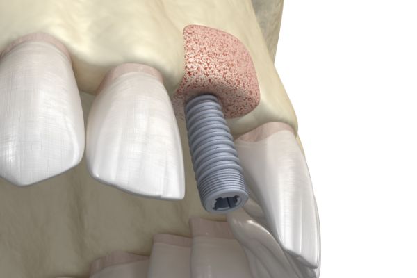
前歯のインプラント治療は、単にインプラントが移植されているだけでは不十分で、インプラントが周囲の歯や歯茎と審美的に調和していなければなりません。そのため、前歯のインプラント治療は歯茎と、そしてこれを支える骨の移植が併用されることが多く、術者には高度な技術と審美的な治療センスが要求されます。
BEFORE
AFTER
前歯のインプラントは周囲と調和し、左右対称に修復されることが求められます。
審美インプラント治療のリスクと治療保証について
審美インプラント治療のリスクと治療保証について
CASE1
前歯における審美的なインプラント症例
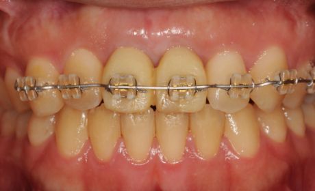
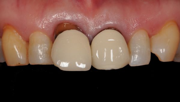
術前の状態
上顎の左側の中切歯の虫歯が大きく、歯根の細菌感染も顕著であったため抜歯してインプラントを行うことになりました。
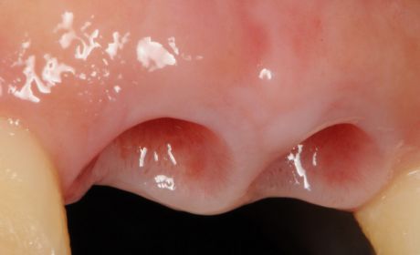
術中の状態
上顎中切歯の抜歯と同時にインプラントを移植し、あわせてインプラント周囲への骨と歯茎の移植を行いました。6ヶ月の治癒期間の後にインプラントと連結するセラミックスの人工歯を作製しました。
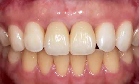
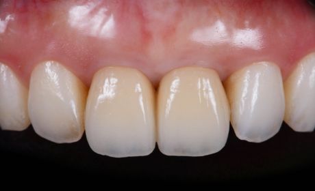
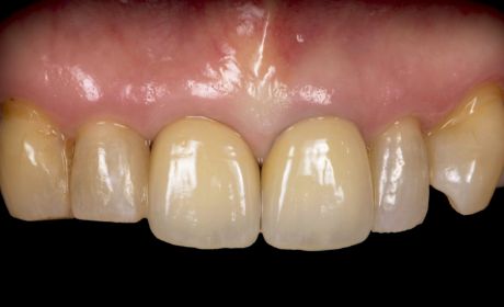
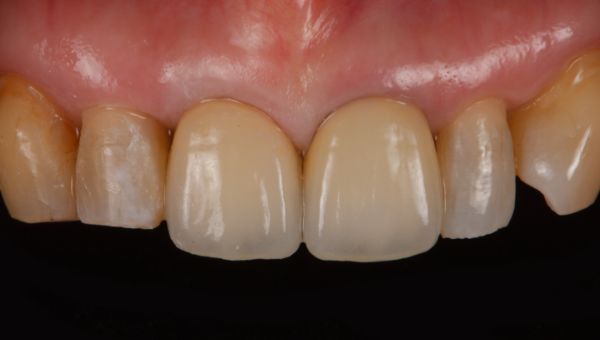
術後の状態
歯のホワイトニングの施術の後に、インプラントと前歯に審美的なセラミックスの人工歯が装着されました。
術前、術後の症例写真
CASE2
前歯だけでなくその周囲の歯茎と骨、
歯並びも改善する複雑なケース
術前の状態
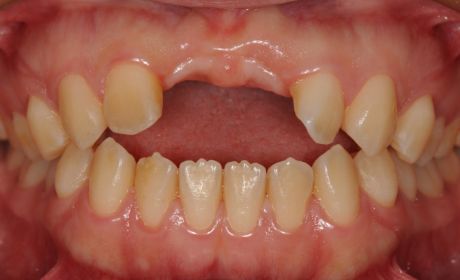
事故により、上顎の前歯と歯茎が失われていました。また、前歯では咬み合わすことができない状態でした(開咬:歯列不正の一種)。
術中の状態

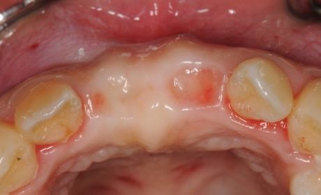
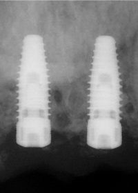
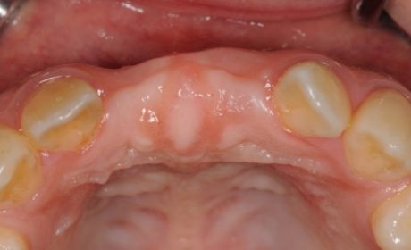
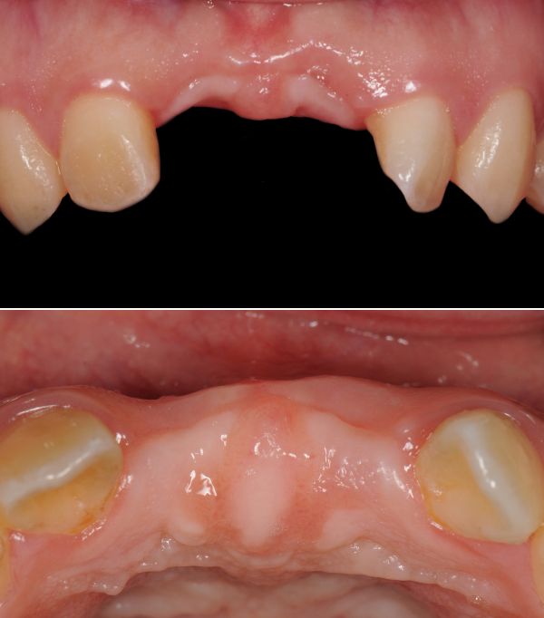
インプラントを支えるための顎骨も失われていたため(写真1)、事前に骨造成術を行い、十分な治癒期間の後に(写真2)、インプラントが移植されました(写真3)。
歯並びの矯正治療が行われ、その後、歯茎の移植も行い歯と歯茎の左右対称性を達成することができました。2本のインプラントを支台とするセラミックスの歯が装着されました。
術後の状態
術後の口腔内所見とX線画像との重ね合わせ。歯だけではなく、骨や歯茎が喪失した症例、あるいは歯列不正の改善のともなう症例では、単にインプラントを移植するだけではなく、様々な治療を融合させ、高次元で審美性を獲得するための高い治療技術が求められます。
術前、術後の症例写真
CASE3
前歯の抜歯と同時に
インプラントが移植されるケース
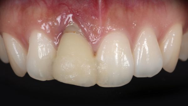
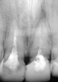
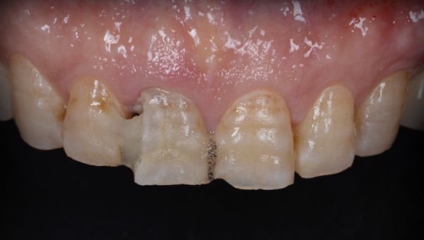
術前の状態
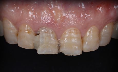
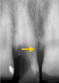
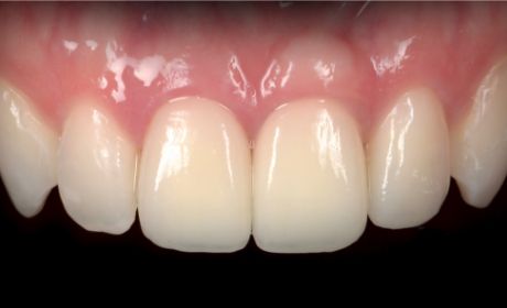
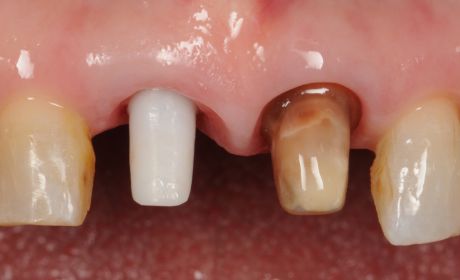
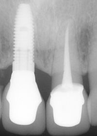
虫歯が重度に進行したため、歯の神経は失活し、歯の破折(写真1矢印)が認められました。本症例では抜歯と同時にインプラントを移植しました。
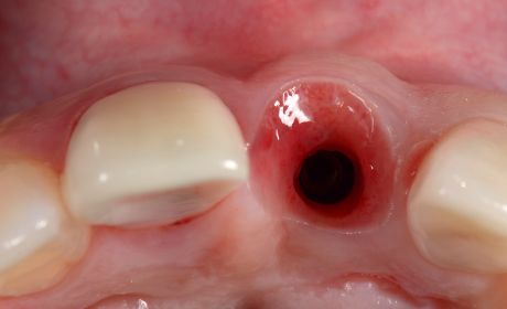
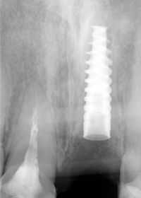
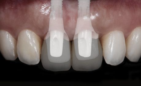
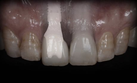
術中の状態
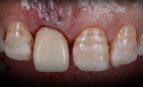
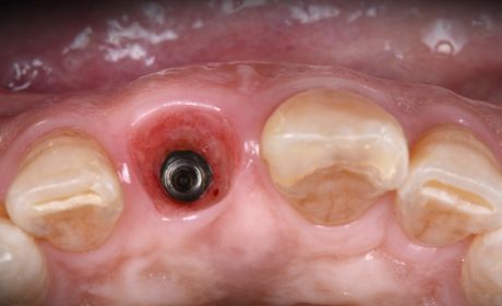
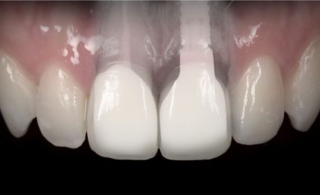
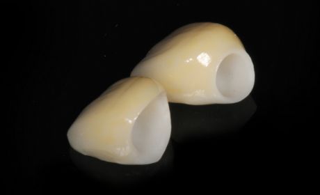
前歯のインプラントは術後に歯茎が退縮する傾向にあるため、これを保証するために骨の移植と歯茎の移植を併用しました。本症例では同時に仮歯も装着され(写真1)、十分な治癒期間の後にセラミックスの人工歯の型取りを行いました(写真2)。
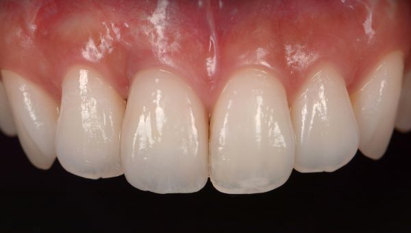
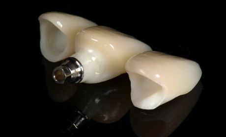
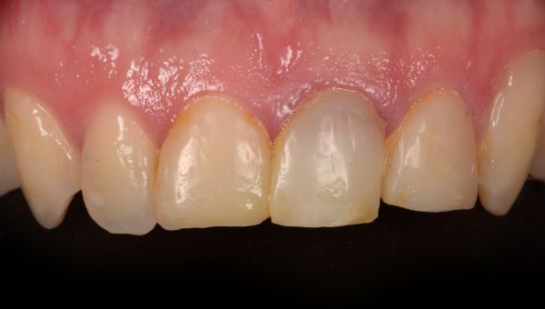
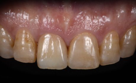
術後の状態
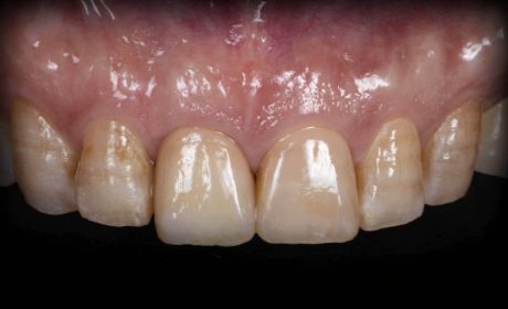
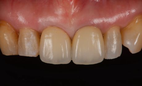
インプラントにセラミックスの歯が装着されました。隣の歯は、歯を一切削らずに樹脂(コンポジットレジン)を用いて修復されました。
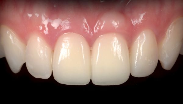
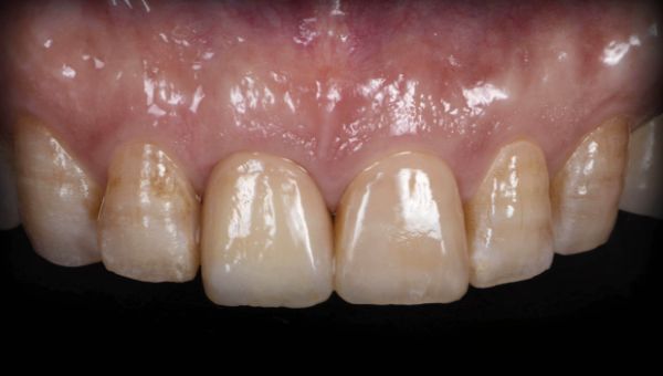
術後3年の状態
インプラント周囲の審美性は変化なく保たれていました。
術前、術後の症例写真
CASE4
歯周病により前歯が抜歯され
インプラントが移植されるケース
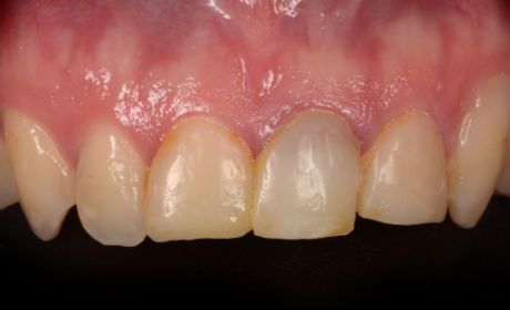
術前の状態
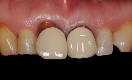
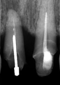
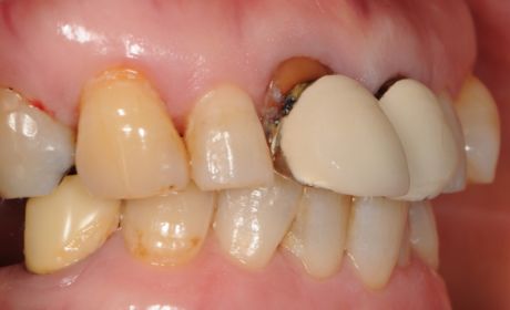
歯周病により前歯が突出し、上顎の右上の中切歯が動揺していました。X線写真にて歯周病が進行していることが認められました。
術中の状態
重度の歯周病では周囲の歯茎が退縮し、歯を支えていた骨も大きく吸収されるため、インプラント治療では骨造成術と歯茎の移植術が必須です。
本症例でも、骨と歯茎の移植術の後に十分な治癒期間を経てインプラントが移植され、審美的なセラミックスの支台とかぶせの歯が作製されました。
本症例でも、骨と歯茎の移植術の後に十分な治癒期間を経てインプラントが移植され、審美的なセラミックスの支台とかぶせの歯が作製されました。
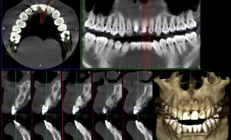
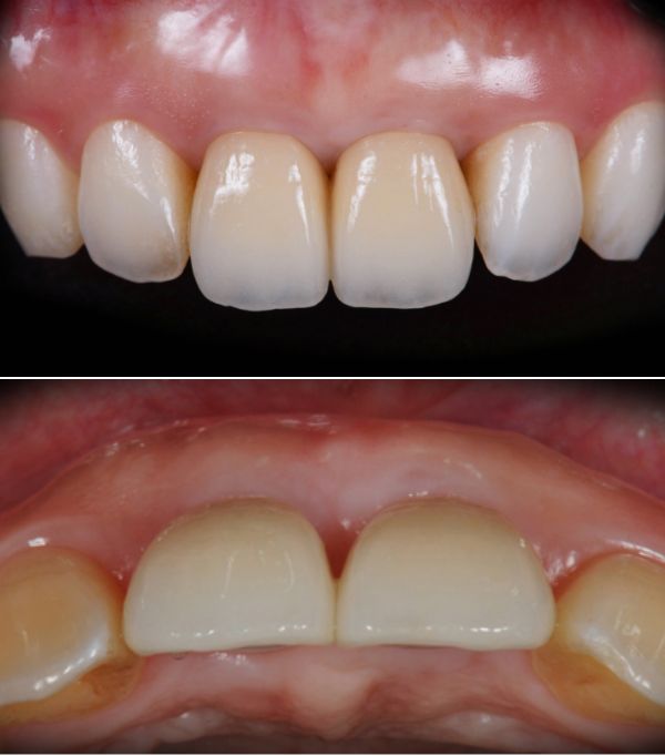
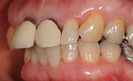
術後の状態
インプラントにセラミックスの人工歯が装着されました。前歯の歯茎と骨が再生されているのが口腔内写真とX線写真から確認できます。
術後8年の状態
インプラントと周囲の歯の審美性は保たれています。
術前、術後の症例写真
前歯のインプラントのリスクと当院の治療保証
前歯のインプラントで歯茎が薄い場合には、術後に周囲の歯茎が退縮し、インプラントを支える骨が吸収する傾向にあります。言い換えると、術後に歯茎の形態が不揃いになり、歯の左右対称性が損なわれるリスクがあります。このため、当院ではインプラント周囲の骨と歯茎の移植術を必ず併用し、歯茎の厚みを増し、インプラントの審美性を長く保つための最大限の配慮を施しております。
また、当院ではインプラント手術、骨造成術、歯肉移植術の初期感染、合併症に対する治療、そして、インプラントが移植後10年の間に脱落した場合の治療費を保証しております。そして、人工歯についても5年間の治療保証を設けております。
また、当院ではインプラント手術、骨造成術、歯肉移植術の初期感染、合併症に対する治療、そして、インプラントが移植後10年の間に脱落した場合の治療費を保証しております。そして、人工歯についても5年間の治療保証を設けております。
免責事項:事故・不注意による外傷、他院での治療・通院、定期検診の通院が途切れた場合には保証対象とならないのでご留意ください。
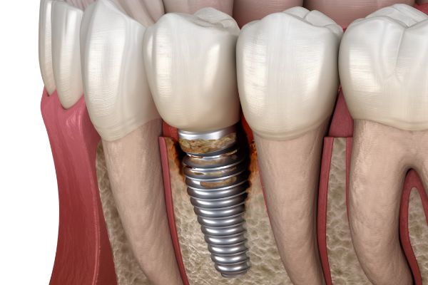
インプラントはご自身の歯と同様に歯周病に罹患することがあります(病名:インプラント周囲炎)。インプラント周囲炎の症状が認められる場合は周囲の歯茎を切開し、インプラント表面を徹底的にクリーニングする手術が必要となります(治療費:4.4万円〜11万円)。